Scanning Electron Microscopy
Imaging and chemical characterization by Scanning Electron Microscopy makes it possible to unravel the identity of trace minerals, the micro-composition of phases, the nature of the micropore systems and the origin of clays. The morphology of the mineral phases and their spatial relationship (particularly of clays) is readily visible by SEM, as well as the spatial pore geometry and flow paths. The X-ray energy dispersive spectrometer (EDX-detector) provides chemical and elemental analysis at any location of interest in the sample, on volumes of only a few cubic micrometers. Specific issues that can be resolved with SEM-EDX (usually in combination with XRD) are:
- Evaluation of formation damage
- Location of damaging clays, zeolites or other mineral phases
- Identification of the nature of any process-induced scales, depositions, sludges, etc. (i.e. non natural samples)
Focussed/Broad Ion Beam – Scanning Electron Microscopy (FIB/BIB-SEM). Where SEM gives a 2-D view of your sample, FIB/BIB-SEM allows to generate a 3-D view of any zone of interest. By ion-milling the sample, the exposed area can be analyzed, layer by layer. It is the ideal tool for the 3D-visualisation of the pore network. It is therefore generally used in the study
of unconventional rock types. Moreover, it allows to:
- Determine the pore size distribution
- Visualize pore connectivity
- Identify the best lithotypes for fracture simulation
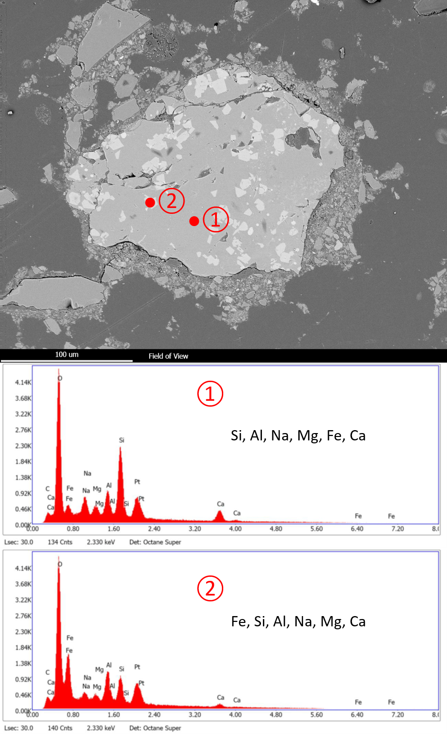
Figure. Example of backscatter electron image of a polished sample showing the presence of an aggregate consisting of a matrix of silicate minerals with Al, Na, Ca, Mg and Fe. The aggregate also contains crystals with a more Fe-rich composition.